

In Vivo Structural Plasticity of Cerebral Vasculature
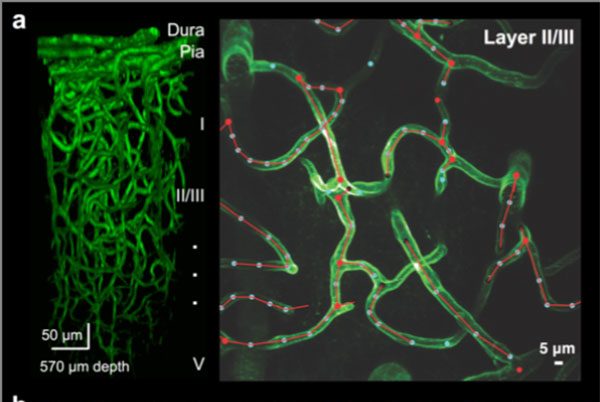
Cerebral vascular networks are stable in the healthy adult. (a) Exemplar in vivo volume of cerebral vasculature in the motor cortex imaged from the dura to deep cortical layers (left), representative image of capillaries within neuronal layer II/III overlaid with 3D tracing (right). Image is a maximal z-projection from 200-220 μm depth (right). (b) In vivo time-lapse images of a descending artery (A), an ascending venuole (V), and capillary segments spanning a one month interval showing stability in vascular networks. Images are maximal z-projection from 100-200 μm depth, scale bar is 10 μm.
- Date and time: March 21, 2017 from 3:30pm-4:30 pm
- Location: Biocomplexity Institute Room 145
- Speaker: Robert H. Cudmore, Ph.D.
- Affiliation: Research Associate. The Johns Hopkins School of Medicine
The cerebral vasculature provides blood flow throughout the brain, and local changes in blood flow are regulated to match the metabolic demands of the active brain regions. This neurovascular coupling is mediated by real-time changes in vessel diameter and depends on the underlying vascular network structure. Neurovascular structure is configured during development by genetic and activity-dependent factors. In adulthood, it can be altered by experiences such as prolonged hypoxia, sensory deprivation and seizure. Here, we have sought to determine whether exercise could alter cerebral vascular structure in the adult mouse. We performed repeated in vivo two-photon imaging in the motor cortex of adult transgenic mice expressing membrane-anchored green fluorescent protein in endothelial cells (tyrosine endothelial kinase 2 receptor (Tie2)-Cre:mTmG). This strategy allows for high-resolution imaging of the vessel walls throughout the lifespan. Vascular structure, as measured by capillary branch point number and position, segment diameter and length remained stable over a time scale of months as did pericyte number and position. Furthermore, we compared the vascular structure before, during, and after periods of voluntary wheel running and found no alterations in these same parameters. In both running and control mice, we observed a low rate of capillary segment subtraction. Interestingly, these rare subtraction events preferentially remove short vascular loops.
For more information, contact Anne Wailes : awailes@vt.edu
